Poster 1
Application of iPSC-derived Mesenchymal Stromal Cell (MSC) toward Treating Muscular Disease
Nana Takenaka-Ninagawa 1, Yuta Itoh 2, Makoto Ikeya 1, Kim Jin Sol 1, Rukia Ikeda 1, Hidetoshi Sakurai 1.
1Center for iPS Cell Research and Application (CiRA), Kyoto University, 2Faculty of Rehabilitation Science, Nagoya Gakuin University, .
+More
Mesenchymal stromal cell (MSC) is well known as a multipotent progenitor for osteogenic, chondrogenic and adipogenic lineage, and is also found in skeletal muscles. Recent studies reported that although they normally act as supportive cells on skeletal muscle homeostasis with producing collagen VI, they are also the responsive cells for fibrosis and fatty degeneration in diseased muscle. Moreover, mutations of Col6 genes, which are specifically expressed in MSC but not in muscle, cause a severe muscle disorder, Ullrich congenital muscular dystrophy (UCMD). Therefore, we assumed that collagen VI positive MSC could be a cell source of cell therapy for UCMD. However MSC isolated from adult tissues are quite limited in expansion. To overcome such hurdle, we plan to use iPSC-derived MSC for future cell therapy of UCMD. In this symposium, I’ll introduce our recent results which demonstrate that human iPSC-derived MSC could ameliorate the phenotype of UCMD model mice (COL6KO/NSG).
Poster 2
Electrical stimulation induces direct reprogramming of dermal fibroblasts into chondrocytes
Gyu Seock Lee 1, Hyuck Joon Kwon 1.
1Department of Physical Therapy and Rehabilitation, College of Health Science, Eulji University, Gyeonggi, Korea
+More
We previously reported that electrical stimulation drives chondrogenesis of mesenchyme stem cells even in the absence of exogenous growth factors. In the previous study, we noticed that electrical stimulation induce MSC chondrogenesis more effectively than chondrogenic growth factors such as TGF betas and BMPs. Therefore, in this study, we have investigated whether electrical stimulation would be able to induce direct reprogramming of dermal fibroblasts into chondrocytes because dermal fibroblast can be easily obtained in comparison to stem cells. We found that electrical stimulation induced human adult dermal fibroblasts to be condensed into the aggregation without addition of exogenous growth factors. qPCR analysis showed that electrical stimulation not only enhanced gene expression of chondrogenic markers such as Col2a1, aggrecan and Sox9, but also reduced gene expression of fibroblast markers such as Col1a1 and Col1a2. Moreover, immunostaining and alcian blue staining also confirmed that electrical stimulation increased significantly expression of Col2 and glycosaminoglycan. These findings would contribute to developing electrotherapeutic strategies for cartilage regeneration. Further studies would be required to elucidate the role of electrical stimulation for cell reprogramming and chondrogenesis.
[Reference]
Hyuck Joon Kwon, Gyu Seock Lee, Honggu Chun., Electrical stimulation drives chondrogenesis of mesenchymal stem cells in the absence of exogenous growth factors. Scientific Reports 6, 39302 (2016)
Poster 3
Research for the appropriate stimulation after mesenchymal stromal cells injection to treat osteochondral defect
Shoki Yamaguchi 1, Tomoki Aoyama 2, Akira Ito 2, Momoko Tanima-Nagai 3, Junichi Tajino 2, Hirotaka Iijima 2, Xiangkai Zhang 2, Wataru Kiyan 2, Hiroshi Kuroki 2
1Department of Physical Therapy, School of Nursing and Rehabilitation Sciences at Odawara, International University of Health and Welfare, Japan
2Department of Physical Therapy, Human Health Sciences, Graduate School of Medicine, Kyoto University, Japan
3Congenital Anomaly Research Center, Graduate School of Medicine, Kyoto University, Japan
+More
The purpose of this study was to research for appropriate follow up treatment after mesenchymal stromal cell (MSC) injection for osteochondral defects in rat knee joint. Allogenic MSCs were injected in the rat knee joint with chronical osteochondral defect. We assessed the effect of exercise and low-intensity pulsed ultrasound (LIPUS) irradiation after MSC injection for the cartilage repair score. MSC injection is effective in stimulating cartilage regeneration on osteochondral defects 4 weeks after injection. Exercise performed after MSC injection could promote cartilage repair in rat osteochondral defect model. On the contrary, LIPUS irradiation after MSCs injection might not promote the cartilage regeneration. Although, the subchondral bone reconstruction was evaluated by measuring the bone volume/ tissue volume in the repaired area of osteochondral defect, LIPUS irradiation might promote subchondral bone reconstruction. This study suggested that exercise and LIPUS intervention might bring the promotion effect of the regeneration in the different tissue. It may become more effective treatment by combining each intervention.
Poster 4
Effects of Mesenchymal Stem Cells In Treating Patients With Knee Osteoarthritis and Role of Rehabilitation –a Systematic Review of Clinical study-
Hirotaka Iijima 1,2, Takuya Isho 1,3, Hiroshi Kuroki 1, Tomoki Aoyama 1
1Department of Physical Therapy, Graduate School of Medicine, Kyoto University, Kyoto, Japan
2Japan Society for the Promotion of Science, Tokyo, Japan
3Rehabilitation Center, Fujioka General Hospital, Gunma, Japan
+More
This systematic review aimed to examine the effects of mesenchymal stem cells (MSCs) in treating patients with knee osteoarthritis (OA) and the role of rehabilitation program on the MSC treatment. Data were collected from randomized controlled trials (RCTs) and non-RCTs that comparing the effects of MSC treatment on clinical symptom and structural outcomes in patients with knee OA. For each electronic database, the endpoint was September 2016. The literature research was performed by two independent examiners. In total, 32 studies were included. Of 32 studies, 23 (71.9 %) studies used MSC intra-articular injection, and the other studies used arthroscopic MSC implantation or those combined with high tibial osteotomy. MSC treatment had high effect sizes in improving knee pain and quality of cartilage, however treatment had a low effect size in increasing cartilage volume. Rehabilitation programs described in the included studies were weight-bearing schedule (37.5 %) and range of motion exercise (28.1 %). However, other information about rehabilitation program such as muscle strength exercise, physical therapy modalities, and preoperative rehabilitation were poor. Although there is an accumulated in vitro evidence that physiological mechanical stimuli promotes MSC differentiation into chondrocyte and extracellular matrix synthesis, rehabilitation program after MSC treatment was insufficient. We believed that establishment of effective rehabilitation protocol is essential in developing regenerative medicine.
Poster 5
Mechanical unloading causes subchondral osteoclast activation resulting in thinning of articular cartilage
Masato Nomura 1, Naoyoshi Sakitani 1, Hiroyuki Iwasawa 1,2, Yoshio Wakimoto 1, Shoko Takano 1, Yuta Kohara 1, Shunsuke Shimaya 1, Hideki Moriyama 1
1Department of Rehabilitation Science, Graduate School of Health Sciences, Kobe University, Japan
2Department of Rehabilitation, St. Marianna University School of Medicine, Japan
+More
Articular cartilage is a highly mechano-responsive tissue. A well-established association exists between mechanical overloading and osteoarthritis, which is characterized by cartilage and subchondral bone alterations. However, the effects of reduced loading on articular cartilage and subchondral bone is still poorly understood. Herein, we investigated the morphological and metabolic responses of articular cartilage and subchondral bone to decreased mechanical stress in vivo, using mouse model of hindlimb unloading by tail suspension. After joint unloading, the thickness of knee cartilage was decreased and subchondral bone atrophy with concomitant marrow expansion predisposed osteoclast activity at bone surface to invade into cartilaginous layer. MMP13 activities were distributed at the bone surface corresponding to the lower edges of articular cartilage. Given that osteoclasts have shown to be capable of expression of MMPs and cartilage matrix resorption, these findings support the concept that the thinning of articular cartilage by mechanical unloading is the result of cartilage matrix degradation and resorption by osteoclasts. Increased RANKL/OPG ratio in chondrocytes at deep zone of cartilage after unloading may partly contribute to subchondral osteoclast activation. Our results should help guide better understanding of crosstalk between articular cartilage and subchondral bone under unloading condition.
Poster 6
Remobilization after 8 weeks of joint immobilization repairs the cartilage surface structure but aggravates site specific degeneration of chondrocyte
Momoko Tanima-Nagai 1, Tomoki Aoyama 2, Hiroshi Kuroki 2
1Congenital Anomaly Research Center, Kyoto University Graduate School of Medicine, Japan
2Department of Physical Therapy, Human Health Sciences, Graduate School of Medicine, Kyoto University, Japan
+More
Within synovial joints, articular cartilage acts as low friction and wear resistant. The pathological changes were reported in animal models of osteoarthritis and joint immobilization including cartilage surface fibrillation or chondrocyte degeneration. These structural alteration lead mechanical, biochemical, cellular and molecular abnormalities in joint tissue. Previous reports using rat immobilization model indicated that joint immobilization causes various degeneration of articular cartilage such as thinning, softening, and surface illegality.
In this study, we investigated that how cartilage changes with remobilization after joint immobilization by using scanning electron microscope (SEM) and transmission electron microscope (TEM). The surface was even and smooth consisted of collagen fibrillar structures were observed in the control group (ctrl) using SEM. After immobilization, the knobby surfaces consisted of a cross-linking fiber network or the leafy and split surfaces were observed at site specifically. With 8 weeks remobilization after then, fiber structure similar to the ctrl was observed again. On the other hand, the degenerated chondrocyte morphology such as cyst formation was observed using TEM. This results showed that the remobilization intervention after immobilization has a negative effect on the chondrocyte at site specific, although has a positive effect on the collagen fiber of cartilage surface
Poster 7
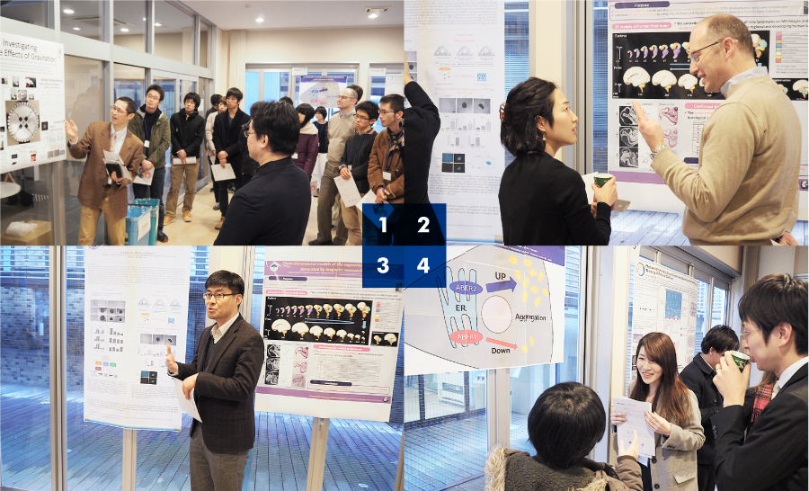
ER Manipulation: A promising therapeutic intervention for protein aggregation diseases
Arisa Yamashita 1, Takamitsu Nakatsuru 1, Hiroki Saito 1, Yuri Hiraki 1, Tetsuo Yamazaki 1
1Department of Molecular Cell Biology and Medicine, Graduate School of Biomedical Sciences, Tokushima University, Japan
+More
Intracellular accumulation of protein aggregates is associated with various neuromuscular degenerative diseases, including amyotrophic lateral sclerosis. At present, no effective therapeutic strategy is available for this spectrum of disease. We recently demonstrated that formation of aberrant protein aggregates can be prevented by tethering αB-crystallin, a chaperone protein, to the endoplasmic reticulum (ER) membrane. To elucidate the underlying mechanisms, we isolated binding partners of the ER-tethered αB-crystallin, and identified two ER transmembrane proteins of unknown function, tentatively designated as ABER1 and ABER2, respectively. ABER1 repressed aggregate formation mediated by the R120G αB-crystallin mutant. In contrast, ABER2 enhanced the aggregation of the mutant. We thus have concluded that the microenvironment surrounding the ER membrane can be a novel therapeutic target to overcome protein aggregation diseases, and that ABER1 and ABER2 are most likely candidates to manipulate to ensure the preventive effects on protein aggregates.
Poster 8
Investigating Multiple Influence of Gravitation
Junichi Tajino 1, Akira Ito 2, Momoko Tanima-Nagai 3, Shoki Yamaguchi 2, Hirotaka Iijima 2, Wataru Kiyan 2, Tomoki Aoyama1 and Hiroshi Kuroki 2
1Department of Development and Rehabilitation of Motor Function, Graduate School of Medicine
2Department of Motor Function Analysis, Graduate School of Medicine, Kyoto University, Kyoto, Japan
3Congenital Anomaly Research Center, Graduate School of Medicine, Kyoto University, Kyoto, Japan
+More
Since every life on Earth has evolved under the Earth’s gravity, altered gravitational stimuli has multiple biological influences. In terms of physical abilities, impairments due to less loading, which are the consequences of inactivity or bedrest are of great interest. Using Hindlimb Unloading and artificial gravity by animal centrifuge, we investigate how various gravitational stimuli affects the physical conditions of rats. As to walking, two-week Hindlimb Unloading evokes gait distortion with their knee and ankle extended throughout the gait cycle, while centrifugation at twice of the normal gravity for 1 hour a day attenuates this alteration. Unloading also diminishes spatial learning ability. In an object-recognition task, unloaded rats show as similar interest in familiar object as new object, which supposed to draw more attention. That implies they do not learn from the environment. Meanwhile, centrifugation ameliorates this learning deficits. However, the benefits of centrifugation might be restricted within a certain range of intensity. Centrifugation at 2.5 times of the normal gravity results less preventive effects from those alterations. Further, intermittent centrifugation affects a circadian rhythm of activity and causes transient drop of deep body temperature. We will keep these investigations to pursue the effects of gravitational stimuli on physical functions.
Poster 9
Three-dimensional models of the segmented human fetal brain generated by magnetic resonance imaging
Yutaka Yamaguchi 1, Momoko Tanima-Nagai 2, Shigehito Yamada 1,2
1Human Health Sciences, Kyoto University Graduate School of Medicine, Kyoto, Japan
2Congenital Anomaly Research Center, Kyoto University Graduate School of Medicine, Kyoto, Japan
+More
The understanding of embryonic development leads to an elucidation of the basic human body structure and congenital anomaly. In recent years, advances in imaging technology have permitted us to obtain more detailed images of the human fetus in a nondestructive and noninvasive manner. However, there is no report that the segmentation is performed for each region of the developing brain using magnetic resonance image (MRI). In this study, we set original landmarks and attempted to create a three-dimensional (3D) models of the developing brain which were reconstructed using MRI. Besides, we confirmed the validity of landmark by comparing our results and the several histological serial sections from the Kyoto Collections. We succeeded in registering a maximum of nine landmarks on MR images and reconstructing 19 sequential models of the regionalized developing brain. The same morphological characteristics were observed on both histological serial sections and MRI. These results will be useful for clinical diagnosis of living fetuses in utero and understanding the growth process of each region of the brain.
Poster 10
Postpartum radiographic changes in pelvic morphology and its relation with symptoms of pregnancy-related symphysis pain
Xiang Ji 1, Masaki Takahashi 2, Saori Morino 2, Hirotaka Iijima 1, Mika Ishihara 3, Mirei Kawagoe 1, Yoko Hatanaka 4, Fumiko Umezaki 4, Mamoru Yamashita 4, Tomoki Aoyama 1
1Human Health Sciences, Graduate School of Medicine, Kyoto University, Kyoto, Japan
2School of Science for Open and Environmental Systems, Graduate School of Science and Technology, Keio University, Japan
3Pilates Studio Wohl, Aichi, Japan
4Kishokai Medical Corporation, Aichi, Japan
+More
The etiology of pregnancy-related pubic symphysis pain (PSP) is usually considered as the change in pelvic biomechanics during pregnancy. However, the morphological changes that occur during puerperium remains unknown. Our study investigated the radiographic changes on conventional X-ray films, indicated a ‘closure’ alteration in the pelvic cavity diameter 1-month postpartum with a significant decrease in the distance between bilateral furthest lateral points of acetabular margins (delivery day 239.1 mm vs 1 month postpartum 237.0 mm) and shortened pubic symphysis separation (delivery day 7.9mm vs 1 month postpartum 6.5mm). However, difference in radiographic diameters between groups with and without rapid recovery during postpartum was not clearly evident.
